Viral Fusion Entry Mechanisms: How Viruses Breach Cellular Defenses and Hijack Host Machinery. Discover the Molecular Secrets Behind Viral Invasion Strategies.
- Introduction to Viral Fusion Entry Mechanisms
- Key Steps in the Viral Fusion Process
- Major Classes of Viral Fusion Proteins
- Host Cell Factors Influencing Fusion
- Molecular Triggers and Environmental Cues
- Comparative Analysis: Enveloped vs. Non-Enveloped Viruses
- Implications for Antiviral Drug Development
- Recent Advances and Emerging Research
- Conclusion: Future Directions in Fusion Mechanism Studies
- Sources & References
Introduction to Viral Fusion Entry Mechanisms
Viral fusion entry mechanisms are critical processes by which enveloped viruses penetrate host cell membranes, enabling the delivery of their genetic material into the cytoplasm and initiating infection. This entry is mediated by specialized viral glycoproteins that undergo conformational changes to facilitate the merging of viral and cellular lipid bilayers. The fusion process is highly regulated and can be triggered by various environmental cues, such as receptor binding or exposure to low pH within endosomes. These mechanisms are not only essential for viral infectivity but also represent key targets for antiviral intervention.
There are two principal pathways for viral fusion entry: direct fusion at the plasma membrane and endocytosis followed by fusion within endosomal compartments. The choice of pathway depends on the virus and the host cell type. For example, human immunodeficiency virus (HIV) typically fuses directly with the plasma membrane, while influenza A virus enters cells via endocytosis and fuses with the endosomal membrane upon acidification National Center for Biotechnology Information. The fusion process is orchestrated by viral fusion proteins, such as the hemagglutinin of influenza or the spike protein of coronaviruses, which undergo structural rearrangements to bring the viral and cellular membranes into close proximity, ultimately leading to membrane merger and pore formation Nature Reviews Microbiology.
Understanding viral fusion entry mechanisms is fundamental for the development of fusion inhibitors and vaccines, as these processes are often conserved and essential for viral propagation. Ongoing research continues to elucidate the molecular details of fusion, offering new avenues for therapeutic intervention against a broad spectrum of viral pathogens.
Key Steps in the Viral Fusion Process
The viral fusion entry process is a multistep sequence that enables enveloped viruses to deliver their genetic material into host cells. The process begins with the attachment of viral surface glycoproteins to specific receptors on the host cell membrane, a step that determines host specificity and tropism. Following attachment, conformational changes in the viral fusion proteins are triggered, often by environmental cues such as low pH in endosomes or receptor binding at the cell surface. These changes expose hydrophobic fusion peptides or loops, which insert into the host membrane, anchoring the virus to the cell.
Subsequently, the fusion proteins undergo further structural rearrangements, drawing the viral and cellular membranes into close proximity. This leads to the formation of a hemifusion intermediate, where the outer leaflets of the two membranes merge while the inner leaflets remain distinct. The process culminates in the opening of a fusion pore, allowing the viral nucleocapsid or genome to enter the host cytoplasm. The efficiency and regulation of these steps are critical for successful infection and are tightly controlled by both viral and host factors. Disruption at any stage can prevent viral entry, making these steps attractive targets for antiviral therapeutics National Center for Biotechnology Information; Centers for Disease Control and Prevention.
Major Classes of Viral Fusion Proteins
Viral fusion proteins are specialized molecular machines that mediate the merger of viral and host cell membranes, a critical step for viral entry. These proteins are broadly categorized into three major structural classes—Class I, Class II, and Class III—each with distinct architectures and mechanisms of action.
Class I fusion proteins are characterized by a predominance of α-helical structures and form trimeric spikes on the viral envelope. Upon activation, typically by low pH or receptor binding, they undergo a dramatic conformational change, exposing a fusion peptide that inserts into the host membrane. This class includes the influenza virus hemagglutinin (HA) and HIV-1 envelope glycoprotein (Env) (National Center for Biotechnology Information).
Class II fusion proteins are primarily composed of β-sheet structures and exist as dimers in the pre-fusion state. Activation, often triggered by acidic pH in endosomes, leads to rearrangement into a trimeric post-fusion form. These proteins, such as the E protein of flaviviruses (e.g., dengue and Zika), insert fusion loops into the host membrane rather than a linear peptide (Centers for Disease Control and Prevention).
Class III fusion proteins combine features of both Class I and II, containing both α-helices and β-sheets. They are exemplified by the glycoprotein G of vesicular stomatitis virus (VSV) and herpesvirus gB. These proteins can be triggered by pH changes or receptor interactions and also form trimeric structures during fusion (National Center for Biotechnology Information).
Understanding these classes is crucial for antiviral drug design, as inhibitors can be tailored to block specific conformational changes or interactions unique to each class.
Host Cell Factors Influencing Fusion
Host cell factors play a pivotal role in determining the efficiency and specificity of viral fusion entry mechanisms. The initial step often involves the interaction between viral fusion proteins and specific host cell surface receptors, which can include proteins, glycoproteins, or glycolipids. The presence, density, and conformation of these receptors on the host cell membrane directly influence viral attachment and subsequent fusion. For example, the human immunodeficiency virus (HIV) requires both the CD4 receptor and a co-receptor (CCR5 or CXCR4) for successful membrane fusion and entry into T cells, highlighting the importance of receptor availability and compatibility Centers for Disease Control and Prevention.
Beyond receptor engagement, host cell membrane composition is another critical determinant. The lipid makeup, including cholesterol and sphingolipid content, affects membrane fluidity and curvature, which can either facilitate or hinder the fusion process. Certain viruses, such as influenza, exploit lipid rafts—microdomains rich in cholesterol and sphingolipids—to enhance fusion efficiency National Center for Biotechnology Information.
Additionally, host cell proteases are often required to activate viral fusion proteins through cleavage, as seen with the activation of the influenza hemagglutinin by trypsin-like enzymes. The expression and localization of these proteases can thus restrict or permit viral entry in a tissue-specific manner World Health Organization. Other cellular factors, such as pH regulators and endocytic machinery, further modulate the fusion process, especially for viruses that utilize endosomal entry pathways. Collectively, these host determinants are crucial in shaping viral tropism and pathogenesis.
Molecular Triggers and Environmental Cues
Viral fusion entry mechanisms are tightly regulated by molecular triggers and environmental cues that ensure fusion occurs at the optimal time and location within the host. These triggers are often specific changes in the host cell environment, such as pH shifts, ion concentrations, or receptor engagement, which induce conformational changes in viral fusion proteins. For example, many enveloped viruses, including influenza, utilize the acidic environment of the endosome to activate their fusion machinery. The low pH induces structural rearrangements in the hemagglutinin protein, exposing the fusion peptide and facilitating membrane merger (Centers for Disease Control and Prevention).
Other viruses, such as HIV, rely on receptor and co-receptor binding at the cell surface as the primary trigger. The interaction of the viral envelope glycoprotein gp120 with CD4 and a chemokine co-receptor (CCR5 or CXCR4) on the host cell surface initiates a cascade of conformational changes in gp41, ultimately driving fusion at the plasma membrane (National Institute of Allergy and Infectious Diseases). Additionally, some viruses, like Ebola, require both receptor binding and proteolytic cleavage by host enzymes within endosomes to activate their fusion proteins (Centers for Disease Control and Prevention).
These molecular triggers and environmental cues are critical for viral infectivity and tropism, as they ensure that fusion is restricted to specific cellular compartments or cell types. Understanding these mechanisms provides valuable insights for the development of antiviral strategies targeting the fusion process.
Comparative Analysis: Enveloped vs. Non-Enveloped Viruses
Viral fusion entry mechanisms differ fundamentally between enveloped and non-enveloped viruses, reflecting their distinct structural features and evolutionary strategies. Enveloped viruses possess a lipid bilayer derived from the host cell membrane, which facilitates direct fusion with host cellular membranes. This process is typically mediated by specialized viral glycoproteins that undergo conformational changes upon receptor binding or exposure to acidic environments, enabling the viral envelope to merge with the host membrane and release the viral genome into the cytoplasm. Notable examples include the hemagglutinin of influenza virus and the spike protein of SARS-CoV-2, both of which orchestrate membrane fusion through well-characterized, multi-step processes National Center for Biotechnology Information.
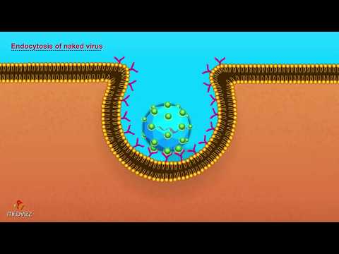
In contrast, non-enveloped viruses lack a lipid envelope and therefore cannot fuse directly with host membranes. Instead, they typically rely on alternative entry strategies such as endocytosis followed by capsid disassembly or pore formation. After internalization, these viruses often exploit endosomal acidification or proteolytic processing to trigger conformational changes in their capsid proteins, leading to the formation of channels or pores through which the viral genome is translocated into the cytosol. For instance, poliovirus and adenovirus utilize such mechanisms to breach the endosomal membrane Centers for Disease Control and Prevention.
The comparative analysis underscores that while both virus types achieve the ultimate goal of genome delivery, enveloped viruses leverage membrane fusion for rapid entry, whereas non-enveloped viruses depend on more complex, multi-step processes involving membrane penetration or disruption. These mechanistic differences have significant implications for antiviral drug development and vaccine design Nature Reviews Microbiology.
Implications for Antiviral Drug Development
Understanding viral fusion entry mechanisms has profound implications for antiviral drug development. Viral fusion is a critical step in the infection process, as it enables the viral genome to access the host cell cytoplasm. By targeting this step, therapeutics can effectively block infection at its earliest stage, potentially reducing viral load and transmission. Several classes of fusion inhibitors have been developed, particularly against viruses such as HIV and influenza, by interfering with the conformational changes in viral fusion proteins or by stabilizing pre-fusion states to prevent membrane merger. For example, the HIV-1 fusion inhibitor enfuvirtide binds to the gp41 subunit, blocking the formation of the six-helix bundle required for membrane fusion (U.S. Food and Drug Administration).
The structural elucidation of viral fusion proteins, often through cryo-electron microscopy and X-ray crystallography, has enabled rational drug design targeting conserved fusion machinery across diverse viral families (RCSB Protein Data Bank). This approach is particularly promising for emerging and re-emerging viruses, such as coronaviruses, where fusion inhibitors could serve as broad-spectrum antivirals. However, challenges remain, including the high mutation rates of viral fusion proteins and the potential for resistance development. Combination therapies, targeting multiple stages of viral entry or using fusion inhibitors alongside other antivirals, are being explored to enhance efficacy and limit resistance (Centers for Disease Control and Prevention).
In summary, insights into viral fusion entry mechanisms are driving the development of innovative antiviral strategies, with the potential to yield both virus-specific and broad-spectrum therapeutics that can be rapidly deployed against current and future viral threats.
Recent Advances and Emerging Research
Recent advances in the study of viral fusion entry mechanisms have significantly deepened our understanding of how enveloped viruses penetrate host cells, revealing new therapeutic targets and strategies. High-resolution structural techniques, such as cryo-electron microscopy and single-molecule fluorescence, have elucidated the dynamic conformational changes of viral fusion proteins during membrane merger. For example, recent studies on the SARS-CoV-2 spike protein have uncovered intermediate states that are critical for membrane fusion, offering potential intervention points for antiviral drugs and vaccines (National Institute of Allergy and Infectious Diseases).
Emerging research has also highlighted the role of host cell factors in modulating viral entry. Lipid composition, membrane curvature, and the presence of specific receptors or co-factors can dramatically influence the efficiency and pathway of viral fusion. Novel findings suggest that some viruses exploit endosomal acidification or protease activation to trigger fusion, while others can fuse directly at the plasma membrane, depending on the cellular context (Centers for Disease Control and Prevention).
Additionally, the development of advanced in vitro systems, such as organoids and synthetic membranes, has enabled more physiologically relevant studies of viral entry. These platforms facilitate the screening of fusion inhibitors and the dissection of virus-host interactions at unprecedented resolution. Collectively, these advances are paving the way for next-generation antiviral strategies that target the earliest steps of infection (National Institutes of Health).
Conclusion: Future Directions in Fusion Mechanism Studies
The study of viral fusion entry mechanisms has advanced significantly, yet numerous questions remain regarding the precise molecular events and host-pathogen interactions that govern this critical step in viral infection. Future research is poised to benefit from the integration of high-resolution structural biology, single-molecule imaging, and advanced computational modeling to unravel the transient intermediates and conformational changes that occur during membrane fusion. The application of cryo-electron microscopy and time-resolved spectroscopy, for example, promises to capture fleeting fusion intermediates that have eluded traditional techniques (Nature).
Another promising direction involves the exploration of host cell factors that modulate fusion efficiency and specificity. Genome-wide CRISPR screens and proteomics are increasingly being used to identify novel host proteins that interact with viral fusion machinery, offering potential new targets for antiviral intervention (Cell). Additionally, the development of synthetic and reconstituted membrane systems will allow for the dissection of fusion events in controlled environments, facilitating the identification of universal versus virus-specific fusion strategies.
Finally, the ongoing emergence of novel and re-emerging viruses underscores the need for broad-spectrum fusion inhibitors. Rational drug design, informed by detailed mechanistic insights, could yield therapeutics capable of blocking fusion across diverse viral families. As our understanding deepens, interdisciplinary collaboration will be essential to translate mechanistic discoveries into effective antiviral strategies (National Institute of Allergy and Infectious Diseases).
Sources & References
- National Center for Biotechnology Information
- Nature Reviews Microbiology
- Centers for Disease Control and Prevention
- World Health Organization
- National Institute of Allergy and Infectious Diseases
- RCSB Protein Data Bank
- National Institutes of Health